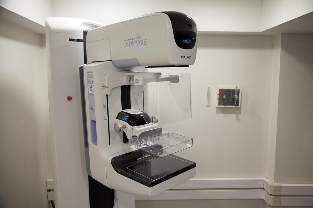
Full Field Digital Mammography
A mammogram is a low-dose x-ray exam of the breasts to look for changes that are not normal. The results are recorded directly into a computer for a doctor called a radiologist to examine. A mammogram allows the doctor to have a closer look for changes in breast tissue that cannot be felt during a breast exam. It is used for women who have no breast complaints and for women who have breast symptoms, such as a change in the shape or size of a breast, a lump, nipple discharge, or pain. Breast changes occur in almost all women. In fact, most of these changes are not cancer and are called “benign,” but only a doctor can know for sure. Breast changes can also happen monthly, due to your menstrual period.
How is a Mammogram Performed?
The patient stands in front of a special x-ray machine. The person who takes the x-rays, called a radiographer, places your breasts, one at a time, between an x-ray plate and a plastic plate. These plates are attached to the x-ray machine and compress the breasts to flatten them. This spreads the breast tissue out to obtain a clearer picture. You will feel pressure on your breast for a few seconds. It may cause you some discomfort; you might feel squeezed or pinched. This feeling only lasts for a few seconds, and flatten your breast, the better the picture. Most often, two pictures are taken of each breast — one from the side and one from above. A screening mammogram takes about 20 minutes from start to finish.
Benefits of digital Mammography
- Long-distance consultations with other doctors may be easier because the images can be shared by computer.
- Slight differences between normal and abnormal tissues may be more easily noted.
- The number of follow-up tests needed may be fewer.
- Fewer repeat images may be needed, reducing exposure to radiation.
- Suitable for younger women with dense breast tissue as well as women aged 50 and over.
The procedure
A mammogram is carried out by a radiographer who will position your breasts on the specially designed mammography machine. In order to obtain a good clear picture, the breast must be held tightly between two pieces of perspex. You may find the scan uncomfortable or painful as the breast tissue needs to be held firmly to ensure a good image is obtained, but this will only last a few seconds. Both front and side images of the breast are taken.
After the scan, you’ll be able to go home immediately.
Please do not use spray deodorant or talcum powder on the day of the mammogram, as this may affect the quality of the X-ray.
Continue taking your normal medication unless you are told otherwise.


