The Worried Well

Welcome to The Imaging Clinic

The Imaging Clinic is committed to ensuring that patients and referring clinicians are confident that they can access the best available diagnostic imaging and image-guided treatments.  We are a long established Chambers of Clinical Radiologists – all Consultants are fully registered with the General Medical Council and with many years of clinical experience.

We offer all major imaging modalities on a number of sites in London and The South East including:

Specialist heart CT Scanning Service with the lowest cost fully reported Cardiac CT Scans in Surrey & The South East from £250.00 for a calcium score. Why not take the scans and reports of our low cost full cardiac package to a cardiologist of your choice anywhere in the world?

Call Us Today For An Appointment
Leading Diagnostic Services In London & The South Of England

The Imaging Clinic has been operating for 25 years and its lead radiologists practicing for between 20-45 years!, offering services to the UK including London, Surrey, Sussex, Hampshire and all the Home Counties.

The clinic co-owns the complex scanning equipment including CT, MRI, Gamma Camera/SPECT and PET CT in joint venture with Circle Mount Alvernia Hospital (Guildford) in joint venture.

Call Us Today for a FREE Consultation

Our Scans & Procedures

The Imaging Clinic offers all major scans and a full reporting service by UK radiologists with many years of training and experience, all registered with the General Medical Council, accredited by The Royal College of Radiologists and with reporting privileges at our hospital base

This technique uses a very low dose of a radioactive agent which is taken up and used by the body’s metabolism to reveal bodily function with the agent broken down naturally by the body usually within minutes to hours before it is excreted.
A technique using a low dose of x-rays to image the entire body in slices as thin as a millimetre demonstrating anatomy and pathology in exquisite detail.
State-of-the-art imaging using a low dose x-ray technique whilst the breast is compressed allowing accurate delineation of microcalcification and soft tissue abnormalities which may suggest breast cancer.  The same equipment utilises an accurate localisation technique called stereotaxis to place a biopsy needle with minimum discomfort in potentially abnormal areas.
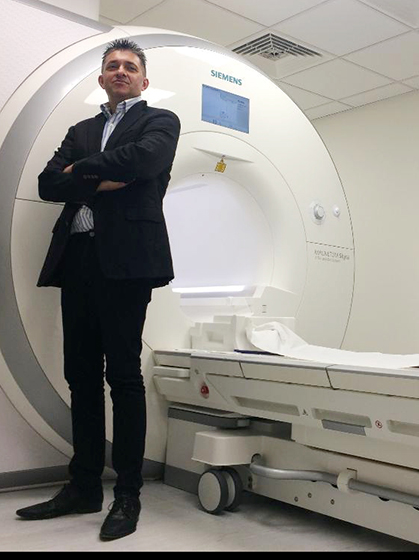
This technique employs a magnetic field  in which naturally occurring particles in the body called ‘protons’ are first ‘excited’ prior to measuring their tendency to ‘relax’.  The measurements are then converted into a detailed image showing anatomy (and pathology where present).
An established ‘minimally invasive’ technique typically using a ‘pinhole’ puncture of the skin at varying sites in the body to allow detailed internal imaging of the body, often used to guided minimally invasive treatments. For a full list of therapeutic options
An infrequently available imaging modality most commonly used to assess tumour activity. A low dose rapidly ‘decaying’ radioactive substance simulating a body metabolite (eg 18F-deoxyglucose [FDG] acting like glucose or PSMA, a biomarker for prostate cancer) is taken up by metabolising cells. Cancer cells typically take up such agents ‘avidly’ and the cancer activity including spread and treatment response can be assessed.
This is a very low dose x-ray technique used to quantitatively measure bone density. The same equipment can also be used to measure the amount and distribution of body fat.
A technique using sound waves rather than x-rays to provide imaging of the body. It is particularly useful in areas of the body where bone or air does not block the penetration of sound waves. Thus it is useful in most areas of the body but limited in the skull, chest and gas-containing structures eg bowel.
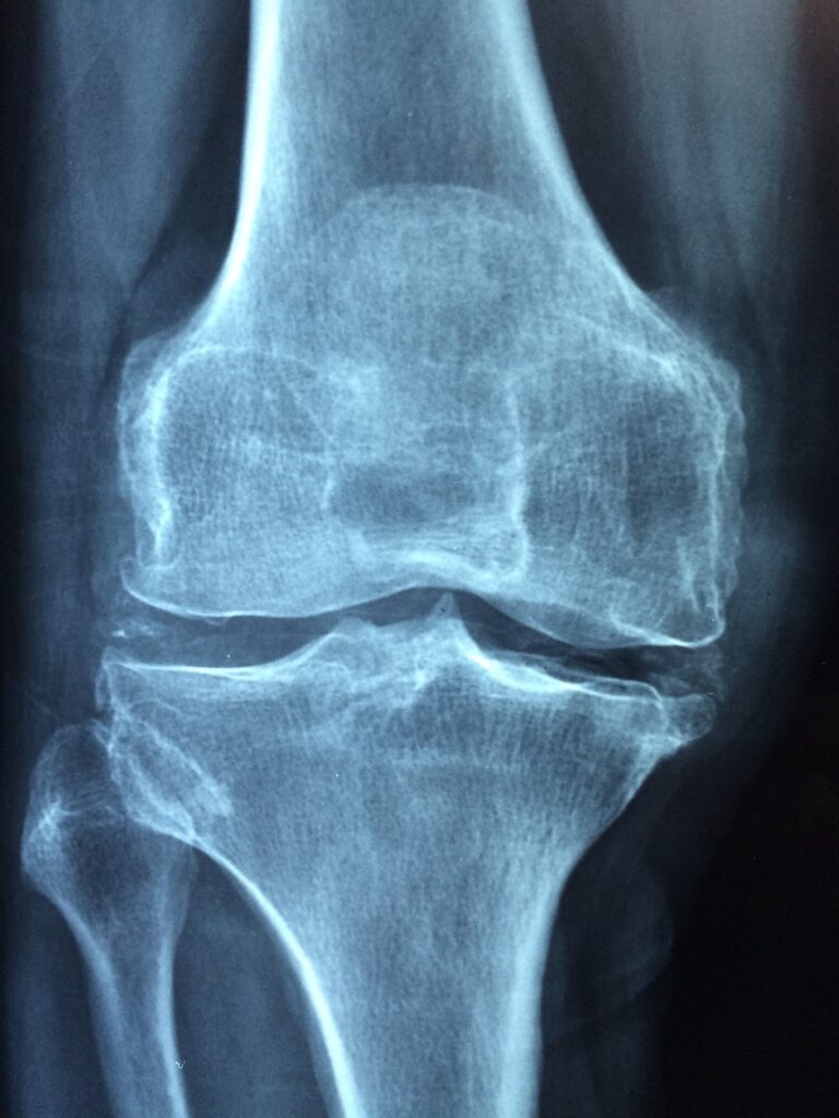
The original imaging technique using ‘x-rays’ following the discovery in 1896 of previously unidentified rays (hence “X”) by Conrad Von Roentgen. Digital imaging using the technique called ‘Computed Radiography’ currently ensures more detailed imaging using lower x-ray doses. The latesrt technique is ‘Digital Radiography’ (DR).

Arrange to see a specialist now

Please contact us for an appointment

Location